02. Heart Physiology
heading
Heart Physiology
ND320 C4 L4 02 Heart Physiology
Recap of ECGs+
Summary
Before we can understand all the parts of the ECG wave, we need to first learn more about how the heart works. The heart is made up of four chambers, two atria and two ventricles. The atria pump blood into the ventricles and then the ventricles pump blood throughout the body. Each heart cell is polarized, meaning there is a different electrical charge inside and outside of the cell. At rest, the inside of the cell is negatively charged compared to the outside. When the cell depolarizes, positive charges outside of the cell flow inside and makes the interior of the cell positively charged relative to the outside. This depolarization causes the cell to contract. The movement of charges across a heart cell’s membrane is the source of the electrical activity that gets measured by an ECG.
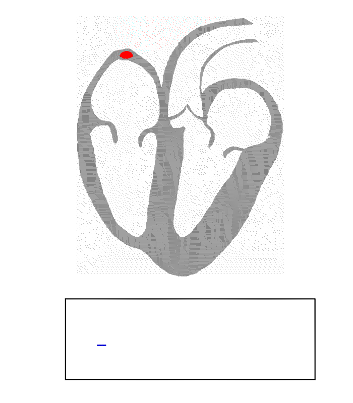
The conduction of electrical impulses through the heart follows a regular pattern orchestrated by the cardiac conduction system. Each heartbeat starts with an impulse in the sinus (SA) node. The sinus node is the natural pacemaker of the heart and is responsible for setting the heart’s natural rhythm. The impulse then propagates throughout the atria and causes the atria to contract. Then, the impulse enters the atrioventricular (AV) node, which delays the propagation of the signal to the ventricles. This gives the atria time to pump blood into the ventricles. After a brief pause, the signal passes through the AV node and into the ventricles, causing the ventricles to contract. This is the largest electrical disturbance in the cardiac cycle. After the ventricles contract, they repolarize, meaning the ions move back across the cell wall to their initial resting state.
Each part of this sequence is responsible for creating a distinct marker-- or wave --in the ECG signal. The P-wave corresponds to the depolarization of the atria. The pause between the P-wave and the Q-wave is caused by the AV node delaying the electrical impulse to the ventricles. When the ventricles contract, we see the large QRS complex. And finally, after some time, the ventricles repolarize, causing the T-wave. The atria repolarize around the same time as the ventricles finish contracting so that activity is not visible in an ECG because it is obscured by the S-wave.
This cycle repeats every heartbeat and results in a very regular and uniform looking signal.
heart animation
Cardiac cycle animation Source: By Kalumet - selbst erstellt = Own work, CC BY-SA 3.0, https://commons.wikimedia.org/w/index.php?curid=438140